Visual electrophysiology can:
- Distinguish between optic nerve diseases, retinal diseases, and functional vision loss
- Identify and monitor damage to the optic nerve
- Examine unexplained vision loss
- Be used as biomarkers for neurodegenerative diseases
Visual electrophysiology in the neuro-ophthalmic practice
Electrophysiology provides objective data on the functionality of the visual pathway from the retina to the visual cortex and back. An evaluation can clearly define the location of visual dysfunction, whether retinal, optic nerve, or post-chiasmal.
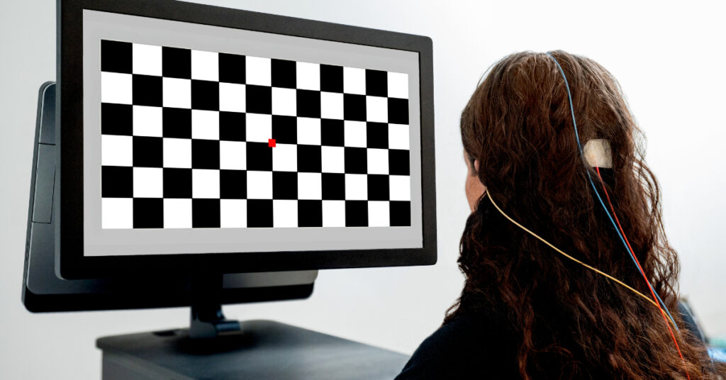
Visually Evoked Potentials (VEP)
VEPs can be used to objectively assess conduction pathway delays caused by compression of the optic nerve, progressive demyelinating diseases, or trauma. They can also be used when optical coherence tomography (OCT) or magnetic resonance imaging (MRI) are inconclusive for confirming conditions like optic neuritis, ischemic optic neuropathy, compressive optic neuropathy, and hereditary optic neuropathies.
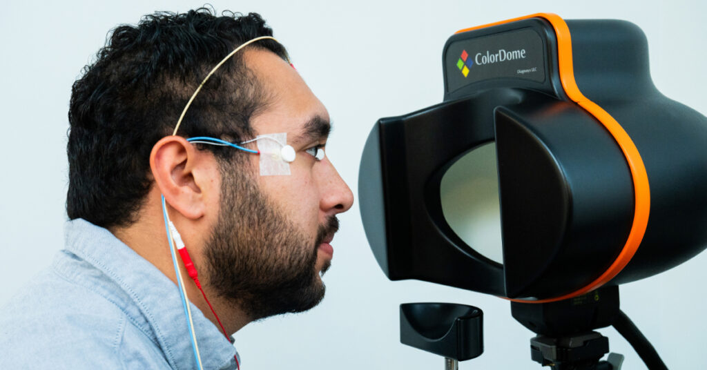
Diagnosys clinical systems support both flash and pattern VEP tests. For pattern tests, you can utilize the LI-LCD monitor while flash VEPs can be performed with either ColorDome® or ColorFlash.
CPT code: 95930
Electroretinography (ERG)
Electroretinography can rule out retinal disorders by isolating the functional integrity of each component of the visual system. This includes photoreceptors, bipolar cells, and inner retinal cells using the general ERG, localized macular dysfunction with mfERG, and retinal ganglion cell damage using PERG or PhNR.
You can perform pattern-based ERGs, such as mfERG and PERG, using the LI-LCD monitor. For flash-based ERGs, like the ERG and PhNR, the ColorDome and ColorFlash stimulators are used. Our Diagnosys clinical systems are compatible with both types of stimulators.
CPT code: 92273
Visual electrophysiology in neurodegenerative diseases
Visually evoked potentials (VEP) are emerging as a non-invasive biomarker for neurodegenerative diseases, offering insights into early detection by identifying characteristic patterns of retinal dysfunction. Patients with conditions like Parkinson’s and Alzheimer’s often experience visual disturbances years before full symptom onset with VEP tests revealing specific changes such as increased latencies in Parkinson’s and altered peak latencies in Alzheimer’s. This capability positions VEP as a valuable tool for early diagnosis and intervention, complementing other biomarkers like cerebrospinal fluid and blood tests.
Explore how VEPs are being used as biomarkers for neurodegenerative disease.