Chiasmal coefficient calculator with a 3-channel visually evoked potential (VEP)
The chiasmal coefficient calculator is a spreadsheet tool that can calculate optic nerve misrouting with the use of a 3-channel Pattern VEP or Flash VEP. Using the raw data of the response, the tool calculates a coefficient and plots a graph. Both of these outputs can be used to draw conclusions about the presence or absence of chiasmal misrouting.
This calculator tool is available at no cost for current Diagnosys system users.
Why would you use this tool?
This chiasmal coefficient calculator may be used as a diagnostic tool for albinism and other misrouting disorders. This method may be used for both children and adults, and can be paired with either a Flash or a Pattern Onset VEP.
Pattern Onset VEPs are preferable. Flash VEPs can be used for patients who are unwilling or unable to fixate, such as young children, or who have low vision or media opacities that prevent effective use of a pattern stimulus.
How to interpret the results?
There are two ways to interpret the results of this chiasmal calculator: graphically or numerically.
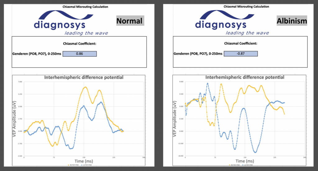
Graphically, the spreadsheet plots the difference between the right and left hemispheric VEP data. In cases of misrouting, the waveforms recorded from right and left eyes will be inverted.
In these graphs, we compare the RE response in yellow and the and LE response in blue. A normal response shows RE and LE data as symmetric. A patient with misrouting will show an inversion of the RE and LE VEPs.

The coefficient values will fall between –1 and +1. Zero indicates pure noise. A negative coefficient value represents an inverse relationship and indicates misrouting. A coefficient closer to +1, indicates symmetrical optic nerve routing.
In the graphs above, the coefficient for the normal patient is 0.86. The patient with misrouting due to albinism has a coefficient of –0.87.
How to use the tool?
The chiasmal calculator is a multi-tabular spreadsheet. Data from any 3-Channel Flash or Pattern VEP may be exported into this tool.
First, set the patient up for a 3-channel VEP, using either the O1 and O2, or PO7 and PO8 electrode locations. Both placements are effective, although Diagnosys recommends PO7 and PO8.
Next, run a Diagnosys-provided 3-channel VEP protocol which calculates the difference between P07-PO8 (or O1 and O2). Ask a Diagnosys representative for this if you do not presently have it available.
Lastly, export your patient’s VEP data and copy/paste it into the spreadsheet. More detailed instructions are provided within the spreadsheet itself. The spreadsheet calculates everything for you, creating a final graph with coefficient.
References
Kruijt CC, de Wit GC, Talsma HE, Schalij-Delfos NE, van Genderen MM. The Detection Of Misrouting In Albinism: Evaluation of Different VEP Procedures in a Heterogeneous Cohort. Invest Ophthalmol Vis Sci. 2019 Sep 3;60(12):3963-3969. doi: 10.1167/iovs.19-27364. PMID: 31560370.
Pott JW, Jansonius NM, Kooijman AC. Chiasmal coefficient of flash and pattern visual evoked potentials for detection of chiasmal misrouting in albinism. Doc Ophthalmol. 2003 Mar;106(2):137-43. doi: 10.1023/a:1022526409674. PMID: 12678278.
Soong F, Levin AV, Westall CA. Comparison of techniques for detecting visually evoked potential asymmetry in albinism. J AAPOS. 2000 Oct;4(5):302-10. doi: 10.1067/mpa.2000.107901. PMID: 11040481.
van Genderen MM, Riemslag FC, Schuil J, Hoeben FP, Stilma JS, Meire FM. Chiasmal misrouting and foveal hypoplasia without albinism. Br J Ophthalmol. 2006 Sep;90(9):1098-102. doi: 10.1136/bjo.2006.091702. Epub 2006 May 17. PMID: 16707527; PMCID: PMC1857410.
