Discover the tests best suited for your patient evaluations
With a system from Diagnosys, you can be assured that you will have the most comprehensive suite of visual electrophysiology tests for your research, clinical trials, or clinical practice. All systems run the same software, which include all the tests defined by ISCEV and more.
Explore the full suite of visual electrophysiology tests
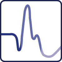
Full-Field Electroretinography (ERG)
Pan-retinal responses to flashes of light in dark and light conditions for diagnosing and monitoring retinal dysfunctions.

Multifocal Electroretinography (mfERG)
Functional assessment of the macula for monitoring the progression of AMD, toxicity, and other conditions.
Pattern Electroretinography (PERG)
Macular ganglion cell response for following up on retinal and optic nerve abnormalities.

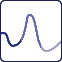
Photopic Negative Response (PhNR)
A pan-retinal test of inner retinal integrity and retinal ganglion cell function for assessing glaucoma and other conditions.
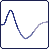
Visually Evoked Potentials (VEP)
Assessment of visual pathway integrity for optic nerve conditions.

Sweep Visually Evoked Potentials (sweep VEP)
Automated, objective assessment of visual acuity for adults and children.
Full-field Stimulus Test (FST)
Psychophysical assessment of a person’s light sensitivity threshold.

Dark Adaptometry (DA)
Psychophysical assessment of photoreceptor sensitivity and time required for dark adaptation.

Electrooculography (EOG)
A measure of the resting potential of the retinal pigment epithelium (RPE) in both darkness and light.