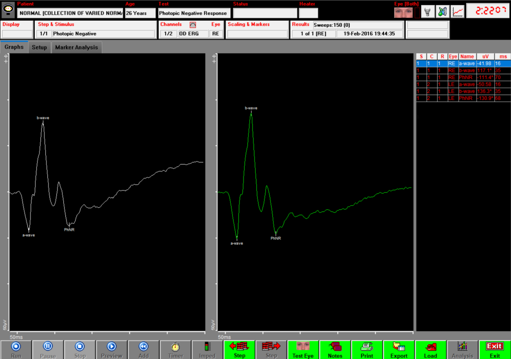
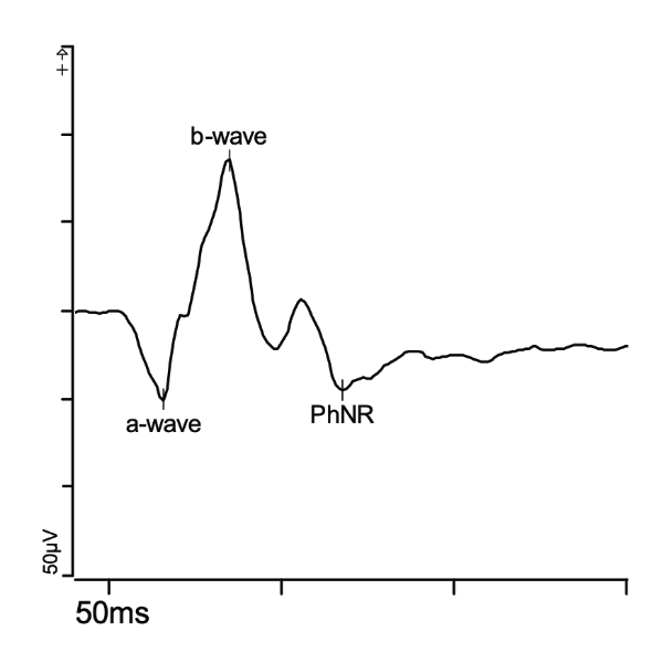
The photopic negative response (PhNR) test is a visual electrophysiology test that can be used to test patients in whom inner retinal integrity and retinal ganglion cell (RGC) function may be compromised. Ganglion cell pathology affects the amplitudes of the PhNR. Reduced amplitudes may be indicators of glaucoma, optic atrophy, central retinal artery occlusion, ischemic optic neuropathy, diabetic retinopathy, or idiopathic intracranial hypertension.

Performing the PhNR test
To complete the PhNR test, a patient sits looking into the ColorDome stimulator with non-invasive corneal electrodes. Flashes of light illuminate the entire visual field while results are recorded and graphed. This test takes under a minute and is often performed alongside the full-field ERG test.
The PhNR stimulus
The PhNR waveform may be visible in the standard full-field ERG test. However, the PhNR test uses a red flash on a blue background in order to generate the largest amplitudes. The most commonly accepted PhNR stimulus is a low intensity red flash on a rod saturating blue background. The ISCEV standard stimulus flashes at 1 Hz. A frequency of 4 Hz can also be used to reduce twitching. Both frequencies are clinically equivalent.

Interpreting PhNR test results
A PhNR test report includes measurements of three key waveforms (a-wave, b-wave, and PhNR) as well as ratios between the PhNR and b-wave. These measurements determine whether the origin of PhNR amplitude change is at the retinal ganglion cells or a more distal retinal location.
References
Frishman, L., Sustar, M., Kremers, J. et al. ISCEV extended protocol for the photopic negative response (PhNR) of the full-field electroretinogram. Doc Ophthalmol 136, 207–211 (2018). https://doi.org/10.1007/s10633-018-9638-x
