Dark Adaptometry is a visual psychophysical test that assesses the sensitivity of photoreceptors and the time required for adaptation to darkness. Prolonged adaptation times can be indicators or conditions such as macular degeneration, diabetic retinopathy, and retinitis pigmentosa.

Perform a dark adaptometry test
The patient sits comfortably in front of the ColorDome, a high performance Ganzfeld stimulator, while holding a single-button response box. When the room lights turn off, an initial bleach phase begins. During this phase, the patient looks into a bright light to bleach the opsins of the retina. After the bleach, the ColorDome‘s lights turn off and the test begins presenting single flashes of light. The patient presses the button each time they see a flash and does nothing if they do not see it. As the test progresses, an algorithm adjusts the light intensity to measure and graph the adaptation curve. An iMask may be added to restrict the field size of the stimulus.
How does dark adaptometry work?
The dark adaptometry test can measure the adaptation of all photoreceptor types using different colors: white, red, green, blue, bluish-green (also known as “rod”), or combinations such as red and green or red and blue. An algorithm embedded in the software automatically chooses the stimulus intensity based on patient responses throughout the test. When the patient consistently responds “yes” to a certain light intensity, the algorithm reduces the brightness of subsequent flashes. Conversely, if the patient indicates they cannot perceive the light, the stimulus intensity increases. This adaptive approach continues throughout the test.
Dark adaptometry output
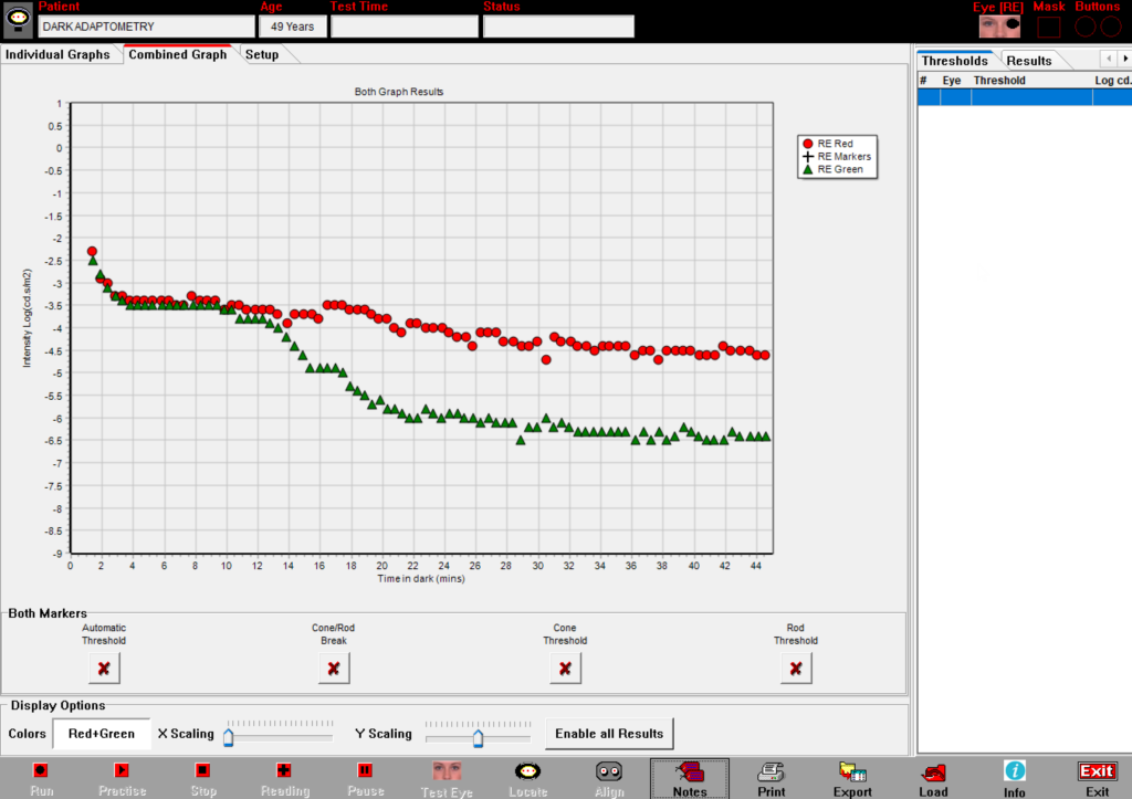
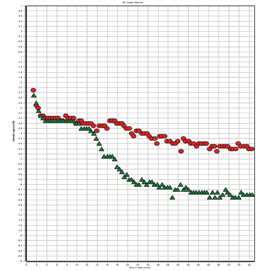
Dark adaptometry tests assess the ability of cones and rods to regain sensitivity after light exposure. The graphs plot luminance on the vertical axis against time on the horizontal axis.

Full rod-cone test
Notice the distinct cone and rod thresholds. Abnormal cone thresholds may indicate cone dystrophy, whereas delayed rod adaptation times could be a sign of age-related macular degeneration (AMD) and abnormal rod thresholds may indicate a rod dystrophy such as Retinitis Pigmentosa.

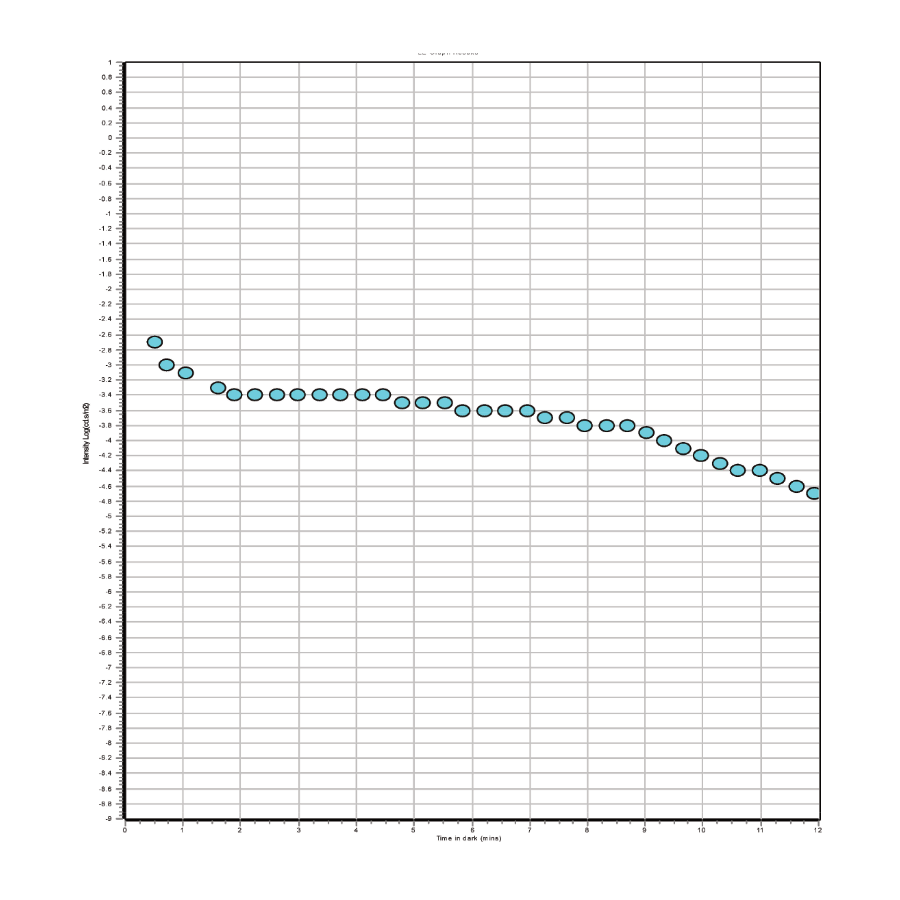
Rod-cone break quick test
This quick test measures the initial phase of rod mediated dark adaptation. Prolonged adaptation times could indicate AMD or diabetic retinopathy, characterized by extended rhodopsin regeneration.

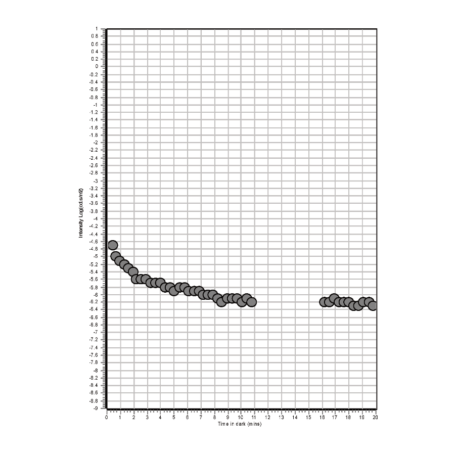
Rod threshold test
The rod threshold test determines the lowest light intensity detectable by the rods, which may provide insights into rod dystrophy.
