Functional Visual Biomarkers
Diagnosys Celeris and Espion products enable precise and easy measurement of specific layers of the retina, including the retinal pigmented epithelium (RPE) and optic nerve.

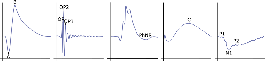
Photoreceptor
Rod and cone responses are primarily isolated in the a-waves of photopic and scotopic ERGs.
Protocols: Scotopic ERG · Photopic ERG · Luminance Intensity Series · Flicker Frequency Series

Bipolar
Bipolar cell activity is primarily reflected in the ERG b-wave under both scotopic and photopic conditions.
Protocols: Photopic ERG · Scotopic ERG

Amacrine
Amacrine cell activity is primarily seen in ERG oscillatory potentials (OPs) and may also contribute to the PhNR response.
Protocols: Scotopic ERG · Photopic ERG · PhNR

Retinal Ganglion Cells
Retinal ganglion cell activity is primarily measured by pattern ERG (PERG) and contributes to the PhNR response.
Protocols: Pattern ERG (PERG) · PhNR

Retinal Pigmented Epithelium
The RPE response is identified by the C-wave component of the ERG.
Protocols: C-wave

Optic Nerve
Visually evoked potentials (VEP) measure signal conduction from the optic nerve to the visual cortex.
Protocols: Pattern VEP · Flash VEP
Flash and pattern stimuli
Celeris offers both flash and pattern stimulation options for rodent and small animal testing.
Flash ERG/VEP
Assess outer and inner retinal pathways with customizable intensities, colors, and durations of light.
Pattern ERG/VEP
Evaluate ganglion cell and cortical responses using contrasting pattern stimuli.
Featured Protocols
These featured examples represent just a few of the many preclinical ERG and VEP protocols available on Diagnosys systems.
Viz Path
Viz Path combines ERG, PhNR, C-wave, and VEP in a single session to evaluate the entire visual pathway, from photoreceptors to visual cortex. This protocol may be useful for studying retinal degenerations, glaucoma, optic neuropathies, RPE dysfunction, demyelinating disease, and therapeutic interventions that target visual function restoration.
Simultaneous Pattern ERG & VEP
The simultaneous ERG and VEP test captures retinal and cortical responses in synchrony. This protocol may be useful for studying optic nerve and visual pathway disorders that include glaucoma, optic neuritis, ischemic optic neuropathy, and demyelinating disease. With additional utility for evaluating neuroprotective or regenerative therapies.

Scotopic Threshold Response (STR)
The scotopic threshold response (STR) measures the lowest detectable retinal responses under dark-adapted conditions, reflecting inner retinal (amacrine and ganglion cell) activity. The STR is valuable for studying early glaucoma, optic nerve injury, retinal ischemia, and post-photoreceptor retinal function in degenerative disease models.

Flicker Frequency Series
A flicker frequency series can evaluate cone-driven retinal function across increasing temporal frequencies to assess signal transmission speed and photopic adaptation. Useful for studying cone dystrophies, diabetic retinopathy, neuroprotective therapies, and pharmacological effects on visual processing.

Preclinical Publications
Diagnosys maintains a list of references to peer-reviewed journal publications that rely on data collected using Celeris®. The papers are classified by animal, test type, and research focus.