Pattern electroretinography (PERG) is a visual electrophysiology test that produces a macular retinal ganglion cell response. When done as a follow-up to an abnormal pattern VEP test, the PERG test can elucidate whether the abnormality is caused by retinal or optic nerve dysfunction. This test may assist in diagnosing glaucoma, optic neuropathies, and primary ganglion cell diseases.
Performing a PERG test
Seat the patient in front of a specialized monitor that can present isoluminant checkerboard patterns. Apply corneal electrodes. The patient fixates on the center of the screen while the checkerboard pattern reverses. This test takes approximately 3 minutes.
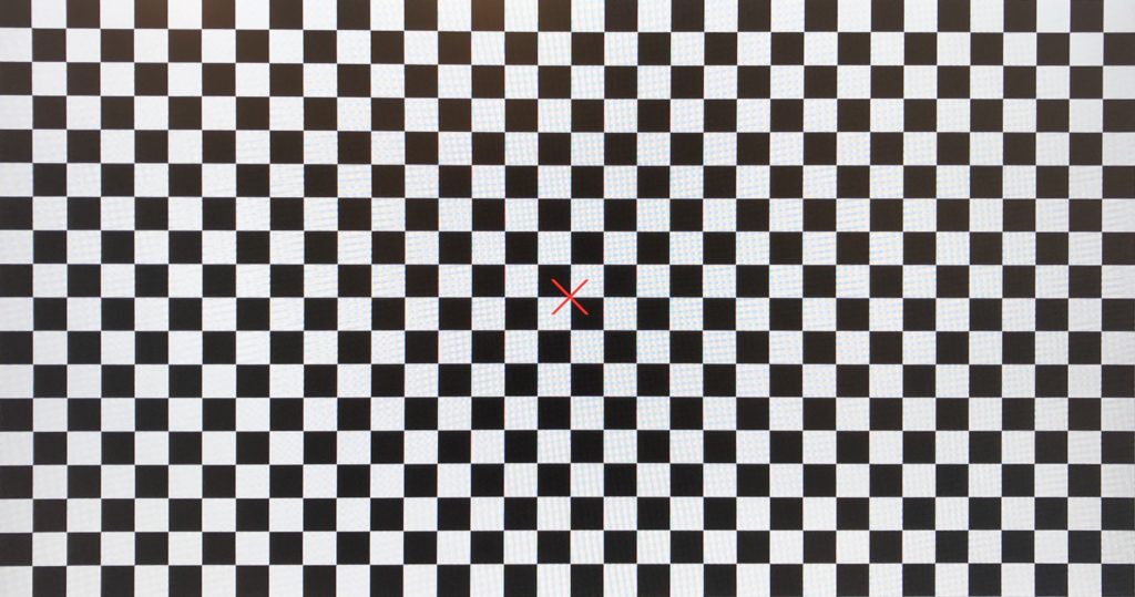
The PERG stimulus
The PERG stimulus is a contrast-reversing, black and white checkerboard pattern much like the pattern VEP. The PERG response is highly sensitive and can be disrupted by flash artifacts. To prevent this, PERGs must be performed using isoluminant displays such as OLED or artifact-free LCD monitors. Monitors with inherent flash artifacts can obscure the PERG signal by stimulating a full-field ERG response on top of the intended PERG response.
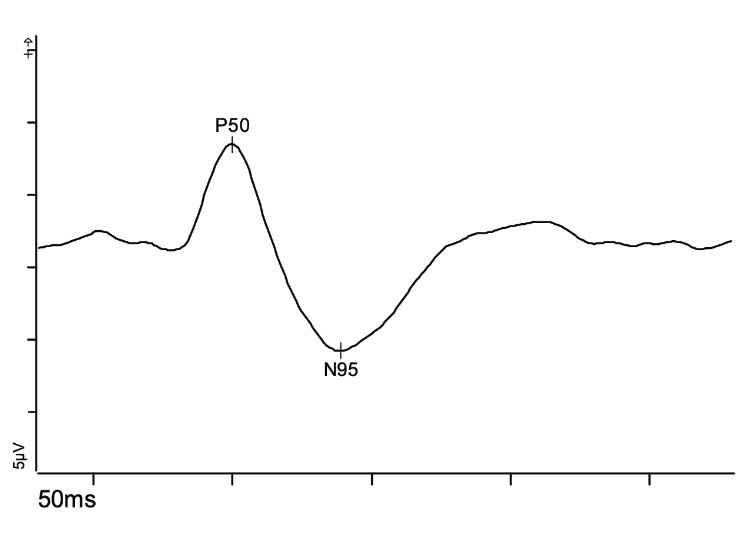
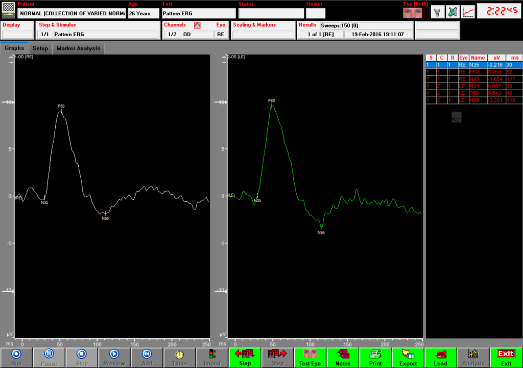
Interpreting PERG test results
The PERG test measures macular cone function. When done as a follow-up to an abnormal pattern VEP, the PERG test can elucidate whether an abnormality is caused by retinal or optic nerve dysfunction. The P50 and N95:P50 ratio both primarily measure macular retinal ganglion cell function and depend upon healthy macular cones. When the P50 alone is reduced or delayed, macular dysfunction is suspected. A reduction of N95 with preservation of P50 suggests dysfunction of retinal ganglion cells.
References
Thompson, D.A., Bach, M., McAnany, J.J. et al. ISCEV standard for clinical pattern electroretinography (2024 update). Doc Ophthalmol 148, 75–85 (2024). https://doi.org/10.1007/s10633-024-09970-1