
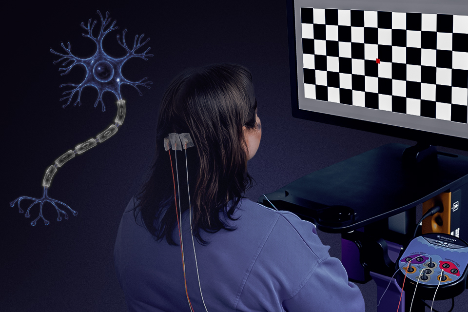
Visual evoked potentials, or VEPs, are increasingly being used in clinical trials for therapies designed to protect, repair, or remyelinate the visual pathway. As multiple sclerosis (MS) research expands from reducing inflammation toward active neural repair, clinical trials will require reliable, objective biomarkers of functional recovery. VEPs assess neural conduction by measuring the timing of signal propagation from the retina to the primary visual cortex, a measurement that is used to determine whether damaged pathways are conducting signals more efficiently than before.
In MS, inflammation damages myelin, the insulating material surrounding nerve fibers. Loss of myelin slows electrical conduction and can affect the optic nerve even when visual symptoms are subtle. Emerging regenerative therapies aim to repair this damage by promoting remyelination, protecting axons, and restoring more efficient neural signaling.
MS clinical trials using VEP as an endpoint
VEP latency reflects the integrity and thickness of the myelin sheath, or conduction velocity. Demyelination typically delays the P100 response, while improved conduction can shorten P100 latency. For this reason, VEP latency has already been used as a clinical trial endpoint in remyelination studies. In the landmark ReBUILD trial of clemastine fumarate, shortening of P100 latency was the primary efficacy endpoint and demonstrated proof-of-concept for remyelinating therapies in chronic demyelinating injury.
This approach continues in newer remyelination studies. The CCMR Two trial evaluated metformin and clemastine with both full-field VEP (ffVEP) P100 latency as its primary outcome and multifocal VEP (mfVEP) latency as a secondary endpoint. Several other ongoing studies are using P100 latency as a primary measure of optic nerve remyelination.
Multifocal VEP (mfVEP) can add another layer by measuring conduction across different regions of the visual field rather than relying on a single global response. In contrast to a standard full-field VEP that can be dominated by surviving, fast-conducting central fibers, the mfVEP can detect regional conduction blocks or localized repair. This can help identify localized delays and may provide additional sensitivity when demyelination affects different portions of the visual pathway unevenly. Research has specifically evaluated mfVEP latency as a potential biomarker for future clinical trials of remyelinating therapies in multiple sclerosis.
Complementing VEP with other test modalities
While the anatomical state of a tissue can be shown by structural imaging such as optical coherence tomoragphy (OCT) for retinal nerve fiber layer thickness and specialized magnetic resonance imaging (MRI) for lesion volume, VEP answers whether the restored tissue can actually transmit functional signals. Combining electrophysiology with structural imaging provides the most comprehensive evaluation of regenerative efficacy in neurodegenerative disease.
As approaches to demyelinating disease move beyond controlling inflammation toward repairing damaged neural pathways, objective measures of conduction become increasingly valuable. VEP offers clinical trials a direct way to measure whether a therapy is translating into improved visual pathway function.