
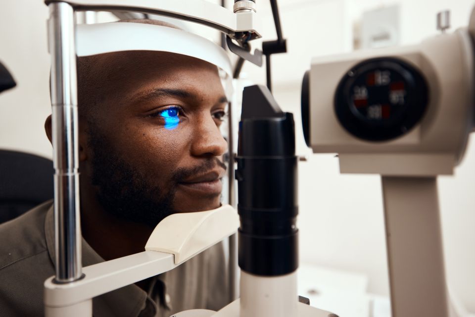
A group from the Center for Ophthalmology, Faculty of Medicine, University of Cologne, Germany, stated in a review paper published in the summer of 2025 that, despite glaucoma being a leading cause of blindness worldwide, there was no consensus on the most effective combination of available test modalities or on the optimal thresholds for glaucoma screening tests. Furthermore, since the onset of glaucoma is largely asymptomatic, it is difficult to detect in the early stages, when it can be easily treated. Still today, a simple, effective, and broadly deployable screening modality for the early stages of glaucoma appears to be an unmet need. Thanks to new studies, visual electrophysiology remains a promising modality for screening for early-onset glaucoma, even as the American Academy of Ophthalmology (AAO) reaffirmed its guidance against routine use in the clinical evaluation of glaucoma.
Diagnosing the early onset of glaucoma is notoriously difficult
Glaucoma is caused by the blockage of the drainage mechanism in the eye that maintains a constant amount of aqueous humor, which the eye produces continuously. A buildup of intraocular pressure (IOP) may indicate an impairment of the aqueous humor drainage mechanism. Understandably, the standard of care continues to rely on gonioscopy and IOP measurements.
High IOP may cause optic nerve damage, preventing retinal signals from reaching the visual cortex and eventually leading to blind spots in the field of vision and, in later stages, total blindness. Standard automated perimetry is widely used to assess visual field impairments caused by glaucoma, but it’s also known to be a long and subjective test. Optical coherence tomography (OCT) is a useful structural imaging modality to help confirm a glaucoma diagnosis by measuring the thickness of the retinal nerve fiber layer (RNFL) and monitoring for thinning over time. OCTs are widely deployed in clinical practice, and retinal structure measurements are relatively quick. Yet, as the AAO points out, measuring RNFL layer thickness is an art. Several reasons can lead to inaccuracies in layer segmentation, including measurement artifacts, differences among equipment manufacturers, and various retinal conditions.
Leveraging visual electrophysiology for glaucoma screening
Visual electrophysiology complements the diagnostic tool set. It can objectively assess the function of retinal ganglion cells (RGCs), which are directly connected to the RNFL. Among the visual electrophysiology suite of tests defined by the International Society for Clinical Electrophysiology of Vision (ISCEV), the pattern electroretinogram (PERG) has long been regarded as a functional test capable of thoroughly assessing the macula and RGCs.
In 2018, the ISCEV published the photopic negative response (PhNR) protocol as an extension of the full-field electroretinography (ERG) test. Like the PERG test, the PhNR test also provides an objective measure of the functional health of RGCs. However, unlike the PERG test, it does not require a test subject to fixate on a target, thereby relaxing the requirements for visual acuity and attention span in the test population. Furthermore, the PhNR test uses a full-field retinal stimulus that provides information about RGCs not only at the center of the retina but also at its periphery, where early glaucoma is likely to manifest, enabling early detection.
The AAO doesn’t recommend visual electrophysiology to diagnose glaucoma
The Glaucoma Panel of the AAO Ophthalmic Technology Assessment Committee (OATC) released a report in June 2026 on the utility of visual electrophysiology testing in the diagnosis of glaucoma. In the report, the panel acknowledged the significant advances in visual electrophysiology’s ability to detect glaucoma accurately. The AAO report recognizes in particular the sensitivity of the PhNR test.
However, based on a review of the literature through August 2025, the panel’s report came short of recommending visual electrophysiology for routine clinical evaluation of glaucoma. The AAO-OATC panel noted that utility is limited by a lack of consensus on stimulation and analysis protocols, including standard reference ranges.
Despite the AAO panel recommendation, we believe that the PhNR test remains a promising modality for glaucoma screening. Recent studies show improvements in the PhNR that should broaden its appeal in clinics.
Improving the sensitivity and specificity of PhNR tests
Promising results from a study evaluating a proposed new PhNR test protocol were published in October 2024. The protocol’s proposed improvements included a systematic method for objectively removing test artifacts caused by eye muscle activity, which adds noise to the measurement.
Improvements were achieved through electrical filtering, a waveform-rejection criterion, and increased stimulus frequency. This modified PhNR test protocol significantly reduced intra- and inter-subject variability in the data, achieving unprecedented sensitivity and specificity.
Administering the PhNR test in a quarter of the time
According to the ISCEV extended protocol, the PhNR test result is conducted with a flash repetition rate of 1 Hz. This allows the PhNR test to recover to baseline. However, a study published in April 2026 demonstrates that the PhNR test can be performed at 4 Hz without compromising test results. By doing so, the authors reduced the PhNR test time from about 200 to 50 seconds. If standardized, this would positively impact clinical workflow and increase its appeal as a screening test, including for glaucoma. And, as the authors note, increasing temporal frequency is particularly important in challenging patient groups. They also point out that higher stimulus frequencies reduce artifacts caused by blinking and eye movements.
Making visual electrophysiology part of the ophthalmologist’s standard toolkit
Visual electrophysiology studies of modified ISCEV test protocols help advance the field. ISCEV, in turn, may help formalize these advances through extended protocols and recommendations. This standardization process is intended to drive clinical adoption, paving the way for routine testing and expanded research. Likewise, recent studies on faster PhNR protocols are promising and could eventually lead to more effective and affordable routine screenings for diseases like glaucoma.