Parkinson’s disease is a progressive neurodegenerative disease mainly seen in elderly individuals that causes neurons in parts of the brain to weaken, become damaged, and die. Neurodegenerative disease biomarkers are an active area of research, some of which focus on visual function. Visual electrophysiology has already demonstrated its capabilities to differentiate between several neurodegenerative diseases and is showing great promise as a biomarker in the early onset of Parkinson’s disease.
From motor symptoms to vision-related biomarkers
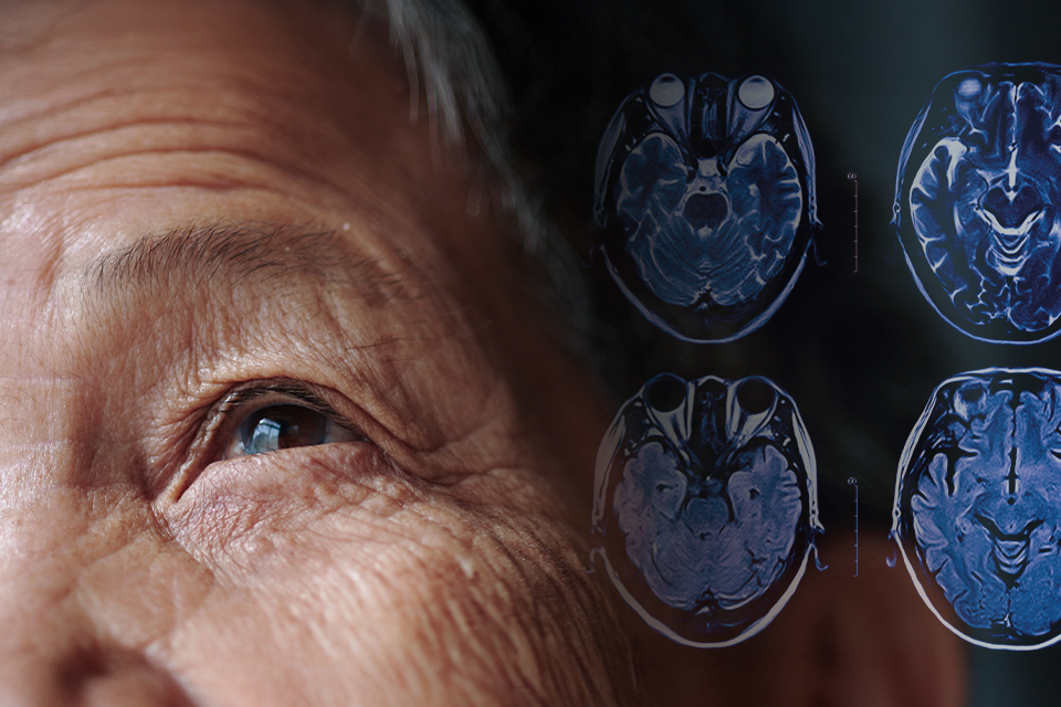
Diagnosing the early stages of Parkinson’s disease could improve patient outcomes. Still, medical practitioners may often find it difficult to distinguish between symptoms of neurodegenerative diseases and other age-related symptoms. One biomarker of neural damage, alpha-synuclein, has been detected in red blood cells, but it is not sufficiently linked to Parkinson’s disease to yield a reliable blood test. Magnetic resonance imaging (MRI) is a proven and reliable diagnostic tool for revealing early stages of neurodegenerative disease. Unfortunately, MRI machines are expensive and not always available.
Today, doctors diagnose Parkinson’s disease and evaluate treatments based on clinical evaluation of motor symptoms. These include tremors, gait characteristics, and resistance to movement. However, some recent research suggests shifting diagnosis and treatment efficacy to measures of the ability to perform activities of daily living, which directly impact quality of life. This, in turn, hinges on adequate visual function.
Approximately 70% of patients with Parkinson’s disease report visual symptoms, and the combination of vision loss, postural instability, and gait disturbances can increase the risk of falls and related injuries. Impairments in visual acuity, contrast sensitivity, and color discrimination are often associated with difficulties performing complex visual tasks. Alpha-synuclein has already been found in the retina of patients with Parkinson’s disease and is known to affect the visual cortex and retinal function. Such findings encourage further studies to identify vision-related diagnostic and prognostic biomarkers of Parkinson’s disease.
Visual electrophysiology biomarkers of Parkinson’s disease
The search for retinal biomarkers of systemic diseases, including neurodegenerative diseases, has often relied on structural retinal images produced by optical coherence tomography (OCT) and other advanced imaging techniques. However, a three-year longitudinal study of a cohort of 114 patients with Parkinson’s disease showed that visual dysfunction is a stronger predictor of outcomes in Parkinson’s disease than retinal thickness.
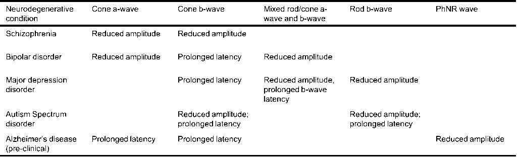
Visual electro-diagnostics, a proven field that follows the standards of the International Society for Clinical Electrophysiology of Vision (ISCEV), offers a wide array of sensitive measures of retinal function that can be useful for research in biomarkers of neurodegenerative diseases. An exciting study recently published a brief review of the electroretinography (ERG) signatures of various neurodegenerative diseases, as summarized in Table 1. This table shows the unique ERG expressions for schizophrenia, bipolar disorder, major depressive disorder, autism spectrum disorder, and Alzheimer’s disease.
Moreover, the paper presented the results of their study on the visual electrophysiology biomarkers of patients with Parkinson’s disease. They observed a delay in the visually evoked potentials (VEP) latency. They also found that scotopic b-wave and PhNR waveforms were impaired in female participants. They indicated that their findings suggest that bipolar cell output is diminished in the rods early in the disease progression, and later in the cones, with retinal ganglion cells ultimately transmitting attenuated signals through the optic nerve. These early changes contrast with more advanced impairments in the later stages of Parkinson’s disease and provide a novel early characterization. The authors believe that these results may lead to a novel biomarker for the early onset of Parkinson’s disease.