While not new, visual electrophysiology for visual neuroscience is a growing field supported by improved instruments, open-source analytical tools, and a new set of variability data to support future preclinical longitudinal studies. This article highlights three papers published in 2025 that showcase this progress.
Research on the retina is relevant to neuroscience
Neuroscience research based on the retina is growing for several compelling reasons. These were extensively reviewed in a recent webinar titled “Visual electrophysiology as a window into the nervous system.” The retina is highly accessible for testing and utilizes a variety of neurotransmitters, including the most common ones. The retina has a well-defined, plastic, and directly visualizable vasculature that responds to perturbations in neural function. It is highly layered, and each layer can be measured using electroretinograms (ERGs). Issues further back in the visual pathway, such as lesions on the optic nerve or strokes, can be evaluated with visually evoked potentials (VEPs). These are mature objective tests that have been standardized by the International Society of Clinical Electrophysiology of Vision (ISCEV).
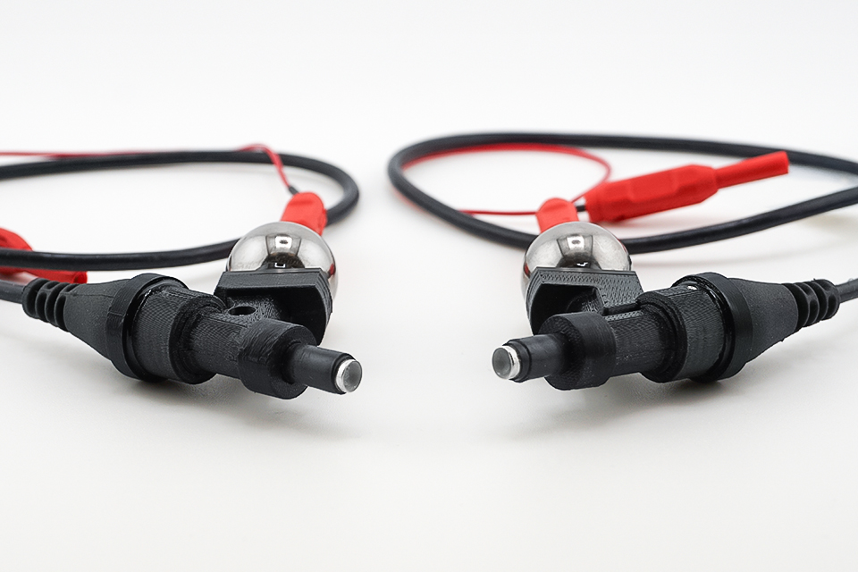
Preclinical visual electrophysiology instruments are improving
Modern ERG and VEP test instruments for small animal models are minimally invasive and designed for high-throughput testing. As noted in a previous article, researchers seeking new preclinical visual electrophysiology equipment will find several highly desirable features, including dual-monocular full-field and pattern stimulators, dichoptic tests, and quick non-invasive setups. The availability of animal-specific stimulators and electrode designs enables efficient testing of a wide range of animals over extended longitudinal studies, from baby mice to adult mice, gerbils, guinea pigs, rats, rabbits, dogs, primates, and pigs.
Open-source statistical software tools
Open-source analytical tools are also evolving, leveraging data export functions developed for preclinical test instruments. A paper published in April 2025 provides an overview of ERGtools2, an open-source visual electrophysiology data processing tool developed in the statistical programming language R. The tool assists neuroscientists in detecting subtle visual electrophysiology trends within noisy data that may provide an early indication of the efficacy of potential new therapies.
Potential biomarker revealed in retinal optogenetics
The author of the ERGtools2 paper published another paper in the same month, this time with colleagues in Germany, the U.K., and the U.S. The team used ERGtools2 and EPhysMethods, another tool developed in R, to reveal an ERG a-wave signature of the interaction between optogenetic prosthetic vision and residual native vision. Specifically, the authors observed an early negative deflection in the ERG signal with sufficient repeatability to be deemed a promising objective measure and potential future biomarker of optogenetic prosthesis contribution to overall vision.
New intra-session repeatability data may support future longitudinal tests
In July 2025, a research team in Melbourne, Australia, published the results of a study on the inter-session repeatability of the ERG and VEP data from the Diagnosys preclinical instrument Celeris. The study was based on 25 healthy wild-type Brown Norway rats. The tests included a comprehensive set of visual electrophysiology measures, including dark-adapted a-wave and b-wave amplitudes, PhNR amplitude, VEP P2 amplitude, and VEP implicit time. All measures of amplitude had a Pearson correlation coefficient between 0.6 and 0.79, a range considered to reflect a “strong” correlation. VEP implicit time showed a “moderate” correlation. The paper also provides the Bland-Altman 95% limits of agreement (LOA) intervals for ERG and VEP parameters, which can be used to design longitudinal studies. In addition, the authors propose a cumulative distribution plot that they believe can reveal statistically meaningful physiological changes.
Advancing visual neuroscience
There are many exciting new developments in neuroscience diagnostic tools, especially in imaging. Visual electrophysiology is an attractive diagnostic tool that provides objective and reproducible data, which promising therapies may rely on to demonstrate their effectiveness. We expect the ecosystem of researchers, developers of analytical tools, and instrument manufacturers to continue building on each other’s momentum to advance visual neuroscience.